
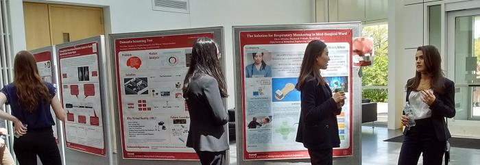
Student Design Projects
The student design project is performed over two semesters. The term "project” is used rather than "thesis" because groups of students generally collaborate on the project, the project can be industrially sponsored, and the project may continue from year-to-year with an evolving group of students. The common sources of projects are the BME faculty, industrial colleagues, and clinicians in the medical and veterinary colleges. The character of different projects is highly variable, e.g., a project might be primarily a theoretical study (e.g., aerodynamics of a neonatal respirator) or might be primarily a laboratory experiment (e.g., tissue engineering of an intervertebral disc).
Project Spotlight: Team Meniscus Repair w/ RF Tissue Welding
Project Spotlight: Pediatric cardiovascular simulator
Project Spotlight: Team BrainLander
Student Team Design Projects
The following is a sampling of recent student team design projects:
Robotic Mobility Device for Toddlers
Click to OpenThe primary method through which toddlers (children 1-3 years old) learn is environmental interaction. This is achieved mainly via independent mobility (i.e., crawling and/or walking). In typically developing toddlers, independent mobility is accompanied by development in emotional, perceptual, cognitive, and social behavior. However, toddlers with independent mobility limiting disabilities have demonstrated apathetic behavior, depressed motivation, and a lack of curiosity and confidence. Providing independent mobility devices may prevent these effects from developing, unfortunately, powered wheelchairs are typically not prescribed to toddlers. Therefore, our team is creating a powered independent mobility device for toddlers with mobility delays/impairments that enables environmental interaction.
External Ventricular and Lumbar Over-Drainage Detection
Click to OpenExternal ventricular drains and lumbar drains (EVDs and LDs) are used to relieve fatal increases in intracranial pressure caused by excess cerebrospinal fluid (CSF). While these drains can be lifesaving, over-drainage of CSF can result in severe neurologic injury or death. Despite the medical necessity and the danger associated with over-draining, these remain one of the few pieces of hospital equipment for which there is no alarm system able to alert clinicians of impending over-drainage. Therefore, our team is creating a non-invasive device that will detect the amount of drained CSF, regardless of color, and alert medical staff when it has exceeded a user-specified threshold.
Detection and Response System for Opioid Overdose
Click to OpenIn the past 12 months, 65,000 Americans have died from opioid overdoses with deaths increasing five fold since 2000. Opioid related deaths are due to over prescription of legal opioids and new, more potent synthetic opioids such as fentanyl flooding the market. Our team has developed a device to treat this epidemic and drastically reduce the number of deaths it causes. Although a treatment exists, it must be administered within a 6 to 8-minute window after onset. As a result, we have designed our device to constantly measure physiological symptoms with a chest belt which triggers our autoinjector to administer the treatment once the user’s biomarkers indicate an overdose.
Liquid Ventilator for Covid-19 patients
Click to OpenIn the COVID-19 pandemic, patients arriving in hospitals sometimes present low oxygen levels however show no trouble breathing, known as silent hypoxia, due to debris found in the alveoli. Effective cleaning of the alveoli is still inefficient through the use of gas ventilation making the ability to clear the alveoli of debris and facilitate gas exchange through a liquid mechanism extremely valuable. This project aims to use PFC (perfluorocarbon) to provide liquid ventilation to patients to reduce the surface tension on alveoli and specifically the pressure when administering oxygen.
Electrical Stimulation Bioreactor
Click to OpenEach year millions of people in the US and across the globe die from heart disease. Therefore it is essential to have the ability to test drugs that can impact the heat in a realistic manner. This requires the usage of mature Cardiomyocytes. This reactor concept is designed in order to promote cardiomyocyte development through the use of mechanical and electrical stimulus. This will result in the development of testable tissues for in vitro drug experimentation.
Uniaxial cell stretcher to measure the biophysical properties of cells and the cell nucleus
Click to OpenGenetic mutations can perturb cellular structure and result in a variety of diseases, ranging from muscular dystrophies to premature aging and cancer. To understand how these mutations affect the mechanical integrity of the cell and its nucleus, it is necessary for a device to induce mechanical strain onto the cells and observe them under in vitro conditions. Currently, the industry standards offer different functionalities, however they do not fully meet our current stakeholder’s needs. This project designed and fabricated a uniaxial stretching device that can apply 20% strain while allowing for high-resolution and live-cell imaging on a confocal microscope.
Craniectomy Prosthesis
Click to OpenOver 100,000 Americans undergo decompressive craniectomies every year, as a result of stroke, infection, or traumatic brain injury. Prevalence of this surgery is exponentially greater worldwide. During the procedure, the bone flap of the skull is removed so that intracranial pressure is reduced but this leaves the surgical site unprotected. Since increasing the time to replace the bone flap minimizes complications, the need exists to deliver skull prosthetics for post craniectomy patients to protect the surgical site. No current solutions address custom surgical site protection. An affordable cranial prosthetic concept is developed to provide optimal protection, comfort, and aesthetic appeal.
Quantifying Physiological and Behavioral Responses to Pain
Click to OpenPain is a highly subjective experience that impacts all people. It is prevalent post-surgery and often associated with medical complications like illness, injury and disability, making it a useful measure for diagnosing and monitoring patients’ ailments. Unfortunately, current pain assessment techniques – Numerical Rating Scale and Visual Analog Scale – have proven faulty and insufficient. These systems rely on self-reporting which is unreliable when patients exhibit drug seeking behavior or are unable to properly communicate their pain. The purpose of this project was to identify physiological and behavioral measures that correlate to pain to improve the pain assessment process.
Augmented Reality for Cancer Mapping
Click to OpenIncreasing recurrence rates after tumor resection surgeries lends to the need for real-time imaging techniques that accurately map cancerous tissue. We modeled a cancerous tumor with BxPC-3 pancreatic cancer cells and used antibody-conjugated microbubbles to serve as a cell-specific contrast agent for US imaging. US images were then post-processed to create 3D reconstructed figures and antibody binding was quantified to model tumor specificity. Future direction includes the use of HoloLens2 to detect the location and distribution properties of tumors in real-time. This enhanced visualization will assist physicians in mapping tumors and in determining optimal treatment options.
Detection of Adverse Events
Click to OpenThe design team focused on developing an approach for the detection and quantification of specific adverse events related to various types of injections. Various approaches to the problem were researched, existing and emerging technologies were compiled and investigated to determine the most promising option. The team narrowed down the detection mechanisms and proposed potential device designs.
Reconstituting Ophthalmic Drugs
Click to OpenThe main purpose of this project is to investigate the effects of different shaking patterns on the successful reconstitution of ophthalmic drugs for veterinary purposes in order to design research protocols. Shaking patterns of ‘gently,’ ‘vigorously,’ and ‘shake well’ were recorded with a MyRIO microcontroller with an accelerometer and analyzed with DIAdem and MATLAB. We have found that 20% of people have the same shake well pattern, and the rest vary greater than one standard deviation.
Uptake Kinetics of Poly(acrylic acid) Microgels
Click to OpenDesign and synthesize poly(acrylic acid) microgels to assess the selectivity of different formulations to absorb and retain targets from an aqueous environment.
Axial Compression Tibial Loading System
Click to OpenIn order to process the Tibial loading on the bond based on a complex mechanical structure in a dynamic environment of bone. As the control demand and experiment setup become highly complex, one of the solutions is to provide a precise control loading system that includes an actuator provided by Zaber.INC and force sensor provided by SENEIT.INC. The software control shall provide the basic flexibility for the user to adjust the loading parameter to match the different types of research experiments’ requirements. The hardware design shall able to hold the mouse knee and feet with designed position properly.
Modeling the Mechanical Behavior of Intervertebral Disc Implants Using Finite Element Analysis
Click to OpenIntervertebral disc degeneration is a complex phenomenon that leads to impairment. Tissue engineering constructs for restoring native tissue require a cage for mechanical stability, however current in vivo results demonstrate cages break in vivo. This highlights a need for a more robust understanding of the mechanics and failure mode. Finite element (FE) analysis allows for biomechanical insight while significantly cutting down on costs and time. We report the development of a FE model that simulates stress localization along the corners of cages under compression between vertebrae. This model provides a method for rapidly testing new cage designs for clinical applications.
Arabidopsis Growth in Martian Regolith
Click to OpenSecuring a sustainable food source will be critical for astronauts and others exploring space in the upcoming years. It is imperative to find a solution that allows space pioneers to grow food and medicinal compounds without adding excessive weight to a space flight. We developed the silica aerogel plant growth medium, the lightest growth media available on the market currently while providing protection against extreme environments. The plants grown in this medium combined with simulated Martian soil grow larger than those grown in regular media. This plant medium optimizes plant growth while minimizing weight when transporting humans in space and granting the ability to thrive beyond earth.
Uniaxial Cell Stretcher to Measure the Biophysical Properties of Cells and the Cell Nucleus
Click to OpenGenetic mutations can perturb cellular structure and result in a variety of diseases, ranging from muscular dystrophies to premature aging and cancer. To understand how these mutations affect the mechanical integrity of the cell and its nucleus, it is necessary for a device to induce mechanical strain onto the cells and observe them under in vitro conditions. Currently, the industry standards offer different functionalities, however they do not fully meet our current stakeholder’s needs. This project designed and fabricated a uniaxial stretching device that can apply 20% strain while allowing for high-resolution and live-cell imaging on a confocal microscope.
AI in the Microelectronics Cleanroom: Label Verification for Large-scale Training Data
Click to OpenMany medical and biological challenges can be solved by Micro Electro Mechanical Systems (MEMS). Such devices are typically fabricated in a microelectronics cleanroom by a sequence of three processes: photolithography, etch, and release etch. Our goal is to use Artificial Intelligence (AI) to improve these processes. AI requires training data with accurate labels but the automatically-generated labels in our database, approximately 100,000 Scanning Electron Microscopy (SEM) images, have errors. In this project, an interactive graphical interface for human verification of the labels named “ImageLabelVoter” was designed, built, and successfully tested.